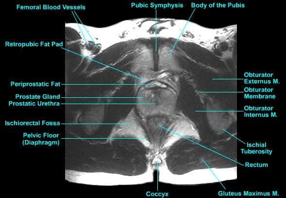
Male Pelvic Floor Mri

Mri Pelvis Anatomy Free Male Pelvis Axial Anatomy
Http Pdf Posterng Netkey At Download Index Php Module Get Pdf By Id Poster Id 117382
Mri Protocols Mri Anatomy Of The Prostrate
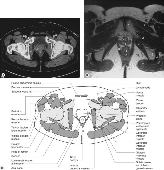
The Pelvis Radiology Key

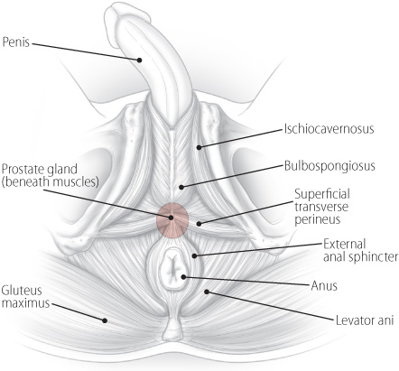
The Pelvic Floor And Male Sexual Function Abdominal Key

Epos C 2394

This mri male pelvis axial cross sectional anatomy tool is absolutely free to use.
Male pelvic floor mri.

Mr Defecating Proctography Radiology Reference Article Radiopaedia Org

Pelvic Anatomy Radiology Human Anatomy

Mri And Us Anatomy Of Female And Male Pelvic Floor Semantic Scholar

Dynamic Mr Imaging Of The Pelvic Floor A Pictorial Review Radiographics

Figure 1 From The Normal Post Surgical Anatomy Of The Male Pelvis Following Radical Prostatectomy As Assessed By Magnetic Resonance Imaging Semantic Scholar

Figure 3 From Morphology And Dynamics Of The Male Pelvic Floor Before And After Retrourethral Transobturator Sling Placement First Insight Using Mri Semantic Scholar

Normal Female And Male Pelvic Anatomy A Axial T2 Weighted Mr Image Download Scientific Diagram
Http Pdf Posterng Netkey At Download Index Php Congress Ecr2014 Module Get Pdf By Id Poster Id 119484

Male Pelvis With Ligaments Pelvic Floor And Organs 7 Parts Includes 3b Smart Anatomy
Pelvic Diaphragm Pelvic Floor

Marilyn Rose Pelvis Ppt Video Online Download

Dynamic Pelvic Floor Mri

About Pelvic Pt Proaxis Therapy Pelvic Pt
Finding Help For Pelvic Pain A Patient S Story Harvard Health
Source : pinterest.com
